

The traditional classification of picoas (Picoa Vittad.) was based on the original descriptions of its two historic species: Vittadini defined Picoa juniperi by its warted surface, while Patouillard described Phaeangium lefebvrei —later combined as Picoa lefebvrei— by a hairy, wartless peridium. To this was later added a criterion widely assumed in the popular literature, namely that spores would be smooth in P. juniperi and warty in P. lefebvrei. However, recent phylogenetic studies have shown that these classic morphological characters do not reliably distinguish the species within the genus.
The first such study, based on ribosomal DNA sequencing —ITS and LSU— and the RPB2 gene from 70 specimens collected across the Mediterranean basin (Zitouni-Haouar et al., 2015, PLOS ONE 10(9): e0138513), revealed that the genus comprises at least six well-differentiated genetic lineages. This study further showed that spore ornamentation is not a reliable diagnostic character, since it may depend, at least in some lineages, on the maturity of the specimen. A second, more recent study (Alvarado, Paz, Lavoise & Van Vooren, 2026, Journal of Fungi 12(2): 84) has considerably expanded the genetic and morphological study of the genus and has proposed recognising at least 19 species within Picoa, 17 of them new to science, grouped into five sections, while also pointing out the existence of further lineages not yet formally described that probably represent additional species.
Thanks to genetic sequencing of our own material, we have been able to identify several of these lineages among the specimens we have collected over the years. However, we do not intend to provide a formal taxonomic description of these species here, since the number of specimens available to us is not sufficient to precisely delimit their morphological variability, especially in relation to the degree of maturity of each specimen. A further, no less important difficulty is that several Picoa species of very similar outward appearance can coexist at the same collecting site; in our case, up to three different species were collected together at a single point, and we were unable to tell them apart in the field. For this reason, we do not consider any specific determination based solely on morphological characters, whether macroscopic or microscopic, to be reliable, and we believe that only genetic sequencing allows each specimen to be confidently assigned to its corresponding species.
In our experience, characters such as the degree of truncation, flattening or central depression of the peridial warts show considerable variability and do not allow the species of Picoa to be reliably separated. These features can be useful for describing the appearance of a particular specimen, but should not be interpreted as reliable diagnostic characters unless supported by molecular data.
Something similar happens with spore ornamentation. In our material, observed under the light microscope, the ascospores appear hyaline, spherical to subglobose or slightly ellipsoidal in outline, with an apparently smooth surface or, at most, finely granulose. Although some recent authors have described more varied spore ornamentation in Picoa, we believe this character should be interpreted with great caution, especially under the light microscope, since it can be affected by the degree of maturity, focus, mounting technique and the perisporium itself. For this reason, we likewise do not regard it as a reliable criterion for species separation when it is not supported by genetic sequencing.
Below we present some of these picoas, all identified from our own sequenced material, together with photographs and locality data. These data are based on our own collections and are not intended to constitute a formal taxonomic revision of the genus, although they document the molecularly confirmed presence of several of these taxa in our study areas. Readers interested in the taxonomy of the genus are referred to the two scientific papers cited above.
Overall, picoas have a peridium ranging from brown to black, and a white gleba that remains so even in fully mature specimens. In young specimens, reddish-brown or ochre mycelial hairs are frequently observed. The asci, quickly evanescent, are subglobose and pedicellate, containing 4-8 globose to broadly ellipsoidal, hyaline ascospores, apparently smooth or weakly ornamented under the light microscope.
In our own collections, and in the mycorrhizae we have studied, picoas always appear associated with the roots of Helianthemum spp. Although some papers cite collections made under or in the presence of Cistus, Tuberaria, Quercus, Pinus or other plants, the presence of these species in the habitat should not be confused with a confirmed mycorrhizal association. Annual Helianthemum plants are often very small and can easily go unnoticed.
Preliminary observations from ongoing studies on the mycorrhizal colonisation of Helianthemum by Picoa species suggest that this genus may have a notably high capacity to form mycorrhizae, which would help explain its frequent occurrence even in areas where no Terfezia fruiting bodies have been detected.
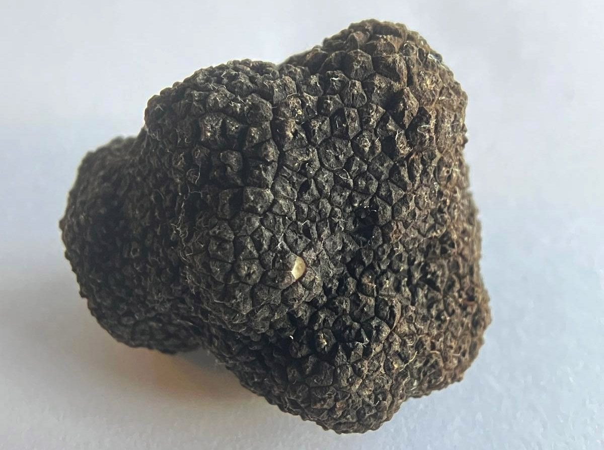
Picoa vazqueziae
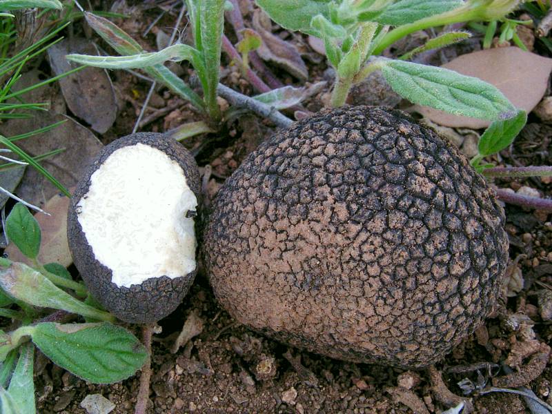
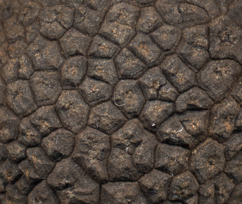
Ascoma subglobose, fairly regular, 1-3 cm in diameter. Peridium black, covered with polygonal, flattened and frequently umbilicate warts, which in some specimens can reach up to 4 mm in width (see detail of the warts). Externally, these specimens closely resemble summer truffles. Gleba white, with white veins enclosing the fertile tissue. Smell sweetish, of coconut. Pleasant taste. Microscopy: asci subglobose, with 6-8 hyaline ascospores, globose to broadly ellipsoidal, with a large guttule, measuring 22-28 (32) x 22-25 (28) µm (Q=1.00-1.16). These ascospores are smooth, and only when mature do they appear finely granulose under the light microscope.
Localities: Casas de Lázaro and Alcaraz (Albacete)
GenBank: JN392150 and OP458213 (both from the same specimen, AH 39035, ITS region, identical in their overlapping portion), correspond to the specimen shown in the photograph, Casas de Lázaro
GenBank: PZ594740, Alcaraz
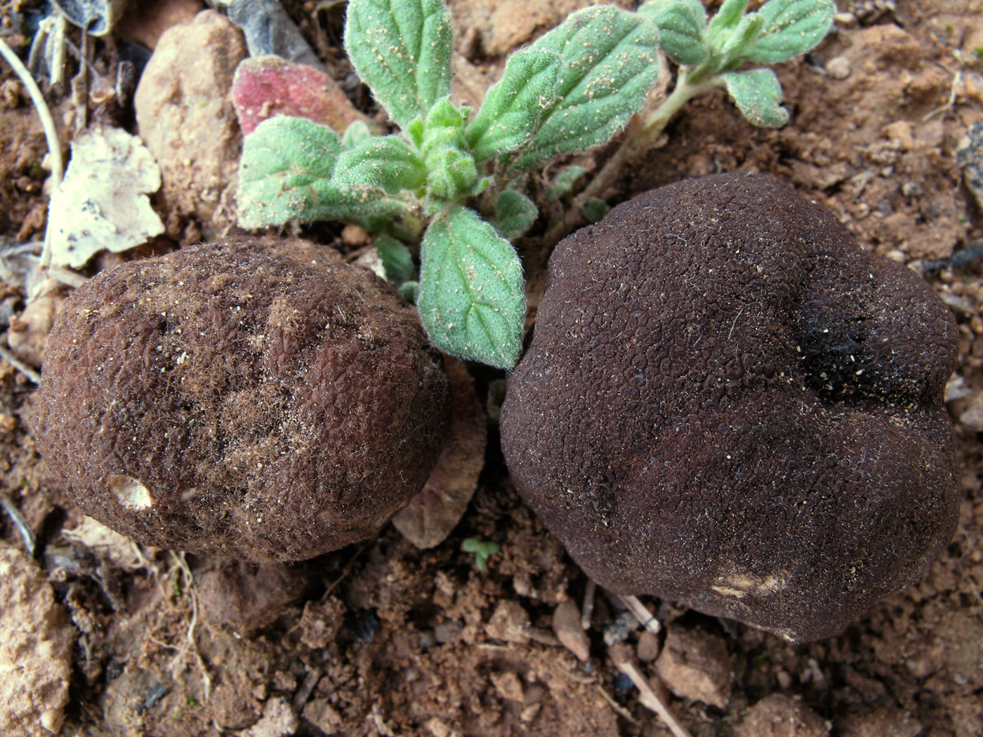
Picoa truncata
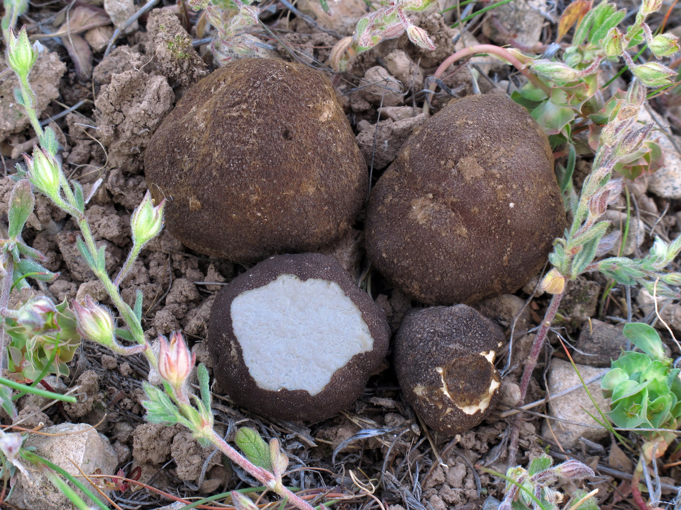
Ascoma subglobose, 1-3 cm in diameter. Peridium verrucose, brown when young and covered with a felt of ochre hairs; at maturity it turns black, with more pronounced warts. Gleba white. These two specimens, apparently so different, were sequenced precisely to confirm that they belonged to the same species. Collecting several picoas at the same site does not imply that they belong to the same species: on occasion we have even collected up to three different Picoa species coinciding in time and place within the same site. Only sequencing allows us to ensure the exact identification of each species.
Localities: Balazote, Chinchilla de Monte-Aragón, Pozuelo (Albacete). Espinardo, Corvera, Águilas (Murcia)
GenBank: PZ594741 (brown specimen in the photograph) and PZ594742 (black specimen in the photograph), Balazote
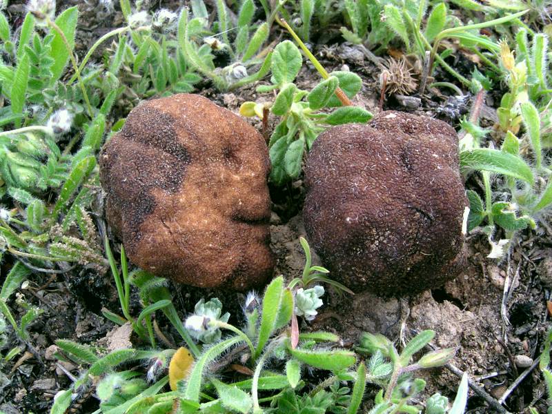
Picoa rodensis
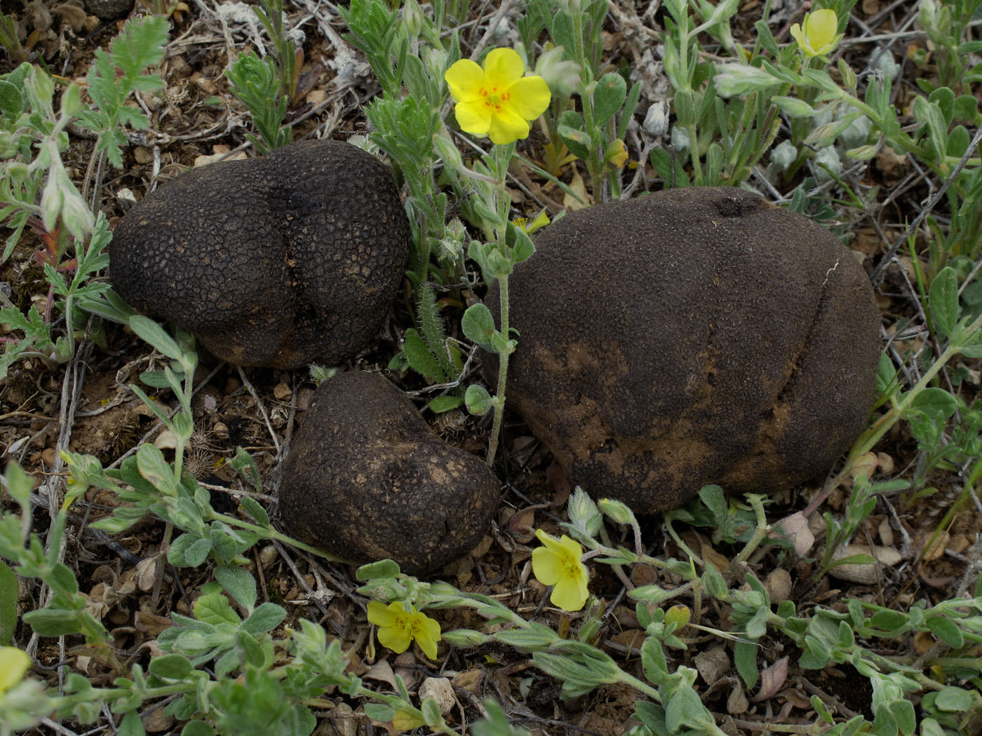
Ascoma subglobose, 2-3 cm in diameter. Peridium black or very dark brown, with strongly pronounced warts. Gleba white.
Localities: La Roda (Albacete). Zarzadilla (Murcia)
GenBank: PZ594743 (corresponds to the specimens shown in the photograph), La Roda
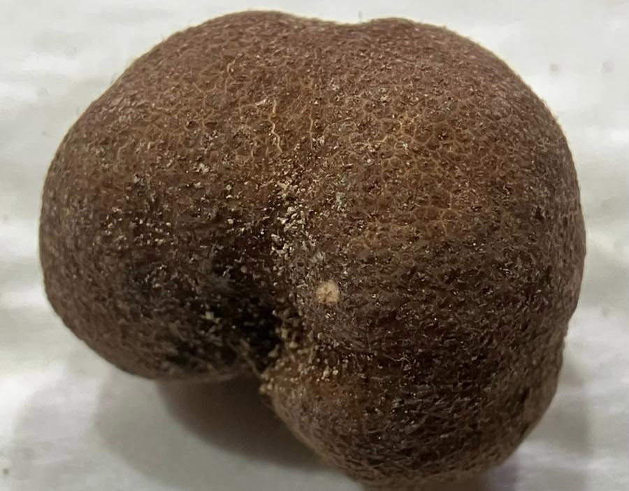
Picoa microspora
Ascoma subglobose to irregularly lobed, 2-3 cm in diameter. Peridium brown to dark brown, with low warts, 1-1.5 mm wide. Young specimens covered with a hairy-looking ochre tomentum, which tends to disappear with maturation or through friction with the substrate. Gleba white. Asci mostly with eight spores. Ascospores not measured. Growing in association with H. canariense. Our Canary Island specimens represent, as far as we know, the first molecular record of this species outside the eastern Mediterranean and North Africa, where it had so far only been known from Egypt, Cyprus and Tunisia.
Localities: Teguise (Lanzarote). Tenerife.
GenBank: PZ594744 (corresponds to the specimen shown in the photograph), Teguise
Picoa fajardoi
Ascoma subglobose to irregularly lobed, 2–3 cm in diameter. Peridium chestnut brown, reddish brown or blackish brown depending on the degree of maturity and moisture.
Localities: Chinchilla (Albacete).
GenBank: PZ670092 (corresponds to the specimen shown in the photograph), Chinchilla
Picoa pontica
Ascoma subglobose-lobed, 2-3 cm, blackish brown, with a strongly warted peridium.
Localities: Torre-Pacheco (Murcia). A frequent species in several localities across the provinces of Murcia and Alicante, confirmed by sequencing (ITS) in 20 collections.
GenBank: PZ670096 (corresponds to the specimen shown in the photograph), Torre-Pacheco, Gallinero del Sur plantation.
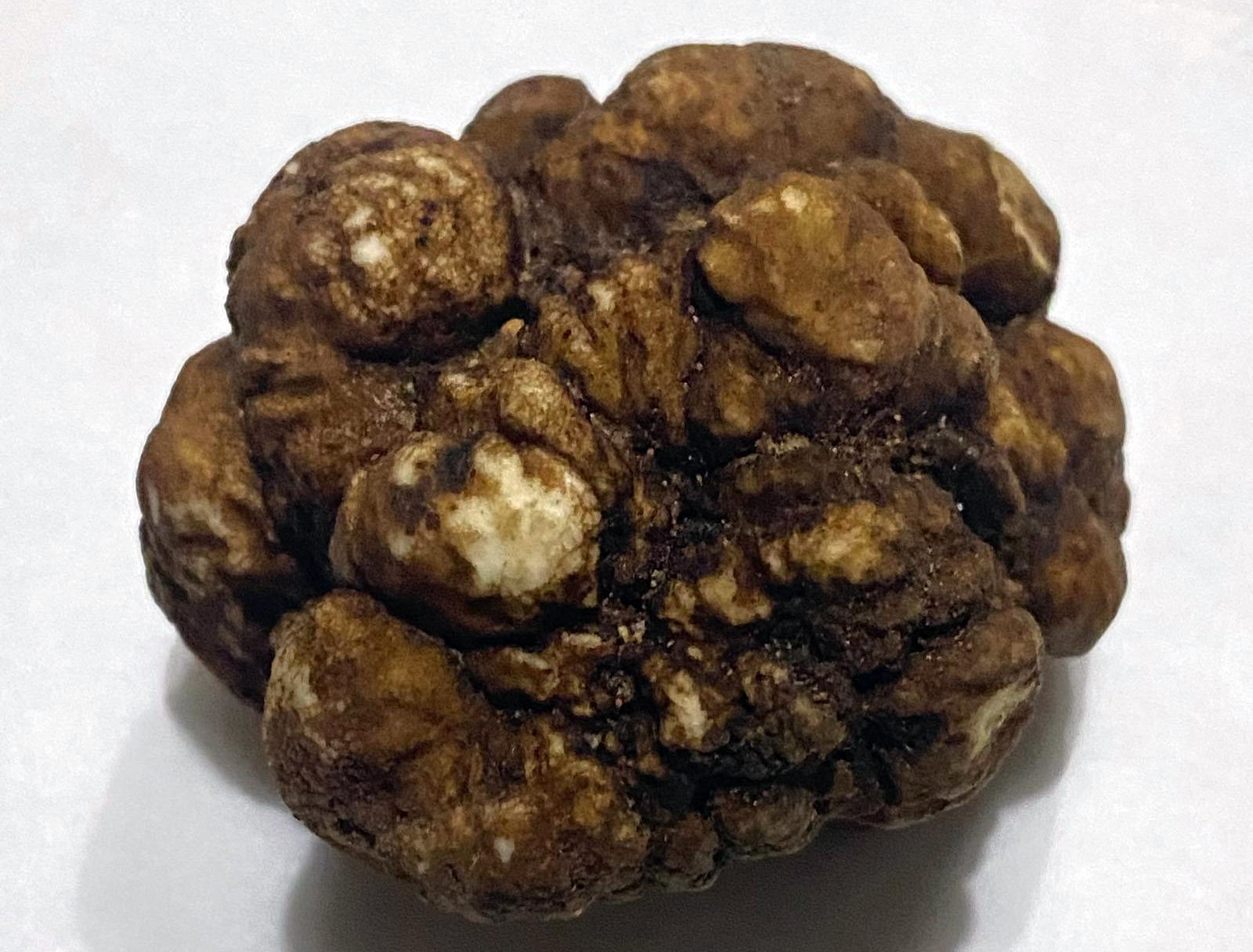
Picoa cartagenensis
Ascoma subglobose to globose, sometimes lobed, 1-5 cm, ranging in colour from orange-brown to a dark, almost blackish brown depending on maturity, with a finely granulose peridium.
Localities: Torre-Pacheco (Murcia).
GenBank: PZ670099 (corresponds to the specimen shown in the photograph), Torre-Pacheco, Gallinero del Sur plantation.
Comments: Picoa cartagenensis is not included in the two general papers on the genus cited in the introduction, as it was described subsequently. The species was published by Moreno, Manjón, López-Saura & Alvarado in Ascomycete.org 18: 85–91 (2026), based on material collected at the Finca El Gallinero del Sur, in a plantation containing specimens of Helianthemum almeriense and H. violaceum previously mycorrhized by the owner of the estate. In that paper it is described as a new species of Picoa sect. Lefebvreorum, based on macro- and micromorphological characters and compared with closely related species of the same section.
Reference: Moreno G., Manjón J.L., López-Saura T. & Alvarado P. 2026. Picoa cartagenensis, a new species of Picoa sect. Lefebvreorum (Pyronemataceae, Pezizales) found in southeastern Spain. Ascomycete.org 18: 85–91. doi: 10.25664/art-0432.
Picoa montecchii
Ascoma subglobose to irregularly lobed, 2–3 cm in diameter, with well-marked pyramidal warts. Peridium reddish brown to dark brown depending on the degree of maturity.
Localities: Torre-Pacheco, Águilas (Murcia).
GenBank: PZ670098 (corresponds to the specimen shown in the photograph), Torre-Pacheco, Gallinero del Sur plantation.
GenBank: PZ670093, Águilas
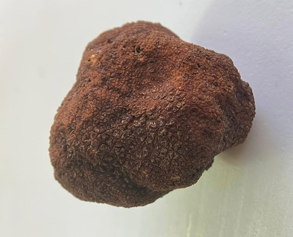
Picoa sp. aff. lefebvrei
Ascoma multilobed, 3 to 5 cm, with rounded lobes and an ochre-brown to chestnut-brown surface that is tuberculate, finely granulose and cracked, showing extensive areas of denuded peridium. It has a markedly different appearance from the rest of the picoas presented here. The collected specimens weighed approximately 20–50 g. This multilobed habit has been observed repeatedly in several collections, suggesting that it is a genuine character of the taxon rather than a one-off artefact of fused primordia.
Localities: Torre-Pacheco (Murcia).
GenBank: PZ670097 (corresponds to the specimen shown in the photograph), Torre-Pacheco, Gallinero del Sur plantation. Our material corresponds to a Picoa that falls molecularly within the P. lefebvrei group. The sequence shows affinity with African sequences deposited in GenBank.
 |
Antonio Rodríguez trufamania@gmail.com antonio@trufamania.com ORCID iD: 0000-0002-0708-7773 |